

DECATUR — From the minute a patient gets the call about an abnormal mammogram, she can go through levels of anxiety: Am I OK? How much is this going to cost? What is the next step?
Physicians understand those worries, and technology is allowing them to use clearer, more precise mammograms to determine whether a patient has breast cancer, reducing false positives and resultant follow-up exams.
Mammography uses low-dose X-rays to detect cancer early, before women experience symptoms and when it is most treatable. Breast cancer is the second most common cancer in women after skin cancer.
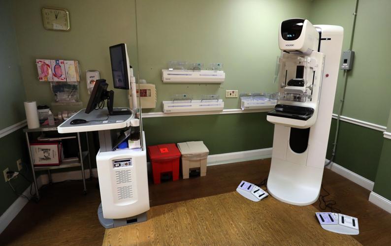
HSHS St. Mary’s Hospital recently began providing breast tomosynthesis, or 3-D mammograms, which allows doctors to view a breast in two and three dimensions.
People are also reading…
“We can see height, width and depth of lesions,” said Daniel Roebein, chairman of the Department of Radiology. “We didn’t have access to that before.”
Decatur Memorial Hospital has offered 3-D mammograms for the past three years.
“It looks like a mammography machine,” said Dr. Jonathan Locke, supervising radiologist for the Decatur Memorial Mammography Center. “You are still clamped in.”
One of the main advantages of 3-D mammograms is the decrease in recall rates. According to Roebein, a recall rate is the number of patients who must return for additional testing.
“If 1,000 people have a screening mammogram, somewhere around 10 percent of those are going to have to have a diagnostic mammogram (a more detailed X-ray),” he said. “A majority of those will not be cancer.”
As described by the National Cancer Institute, screenings with the first mammogram are used to study the breast in patients with no signs or symptoms of cancer.
With the new technology, if a doctor were to recognize an abnormality on the 2-D image, he or she can scroll through the other 3-D images to study the spot. A 3-D mammography uses an X-ray beam to capture more than 100 images on each breast.
“You may see something that looks like a mass, when it is just overlapping tissue,” Roubein said. “In the past, we would have to call the patient back.”
Since 3-D imaging has been available, the recall rate has been reduced by 20 percent, Roubein said. The quality and speed of the 3-D image as well as the low dose of radiation is optimized with the new equipment as well.
According to Locke, 3-D mammograms are especially beneficial for women with dense breast tissue.
“That is the whole focus,” he said.
The latest studies on mammograms showed 2-D exams were not detecting cancers hidden among dense breasts, Locke said.
“Women who have dense breast tissue seem to have a slightly higher risk of breast cancer compared to women with less dense breast tissue,” the American Cancer Society notes on its website. “We do know that dense breast tissue makes it harder for radiologists to see cancer.”
As well as hidden tumors, cancers can be more common in dense breast because of structure of the breast.
“They have more glandular elements, and the cancer comes from the ducts in the glandular elements,” Locke said. They are more at risk for getting breast cancer, and we are worse at finding it with regular mammograms.”
Most insurance plans now include 3-D mammograms.
DMH recommends 3-D mammograms to patients with dense breast tissue. Those with higher risks of breast cancer may receive other exams such as an ultrasound or MRI.
According to Locke, breast tomosynthesis reduces false positives for patients, resulting in less radiation as well as follow-up exams, medical costs and anxiety.
For those who have 2-D mammograms, the process is still useful at detecting abnormalities that might require further diagnosis from the doctor.
“It is not substandard,” Roubein said. “It is very reliable.”
At St. Mary’s, patients receive both a 2-D and 3-D mammogram at the same time. The 3-D procedure is similar to a standard mammograms with a few extra seconds added.
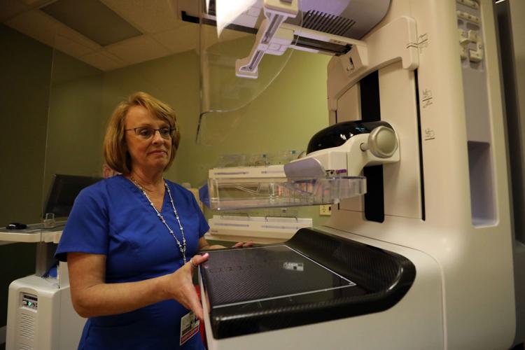
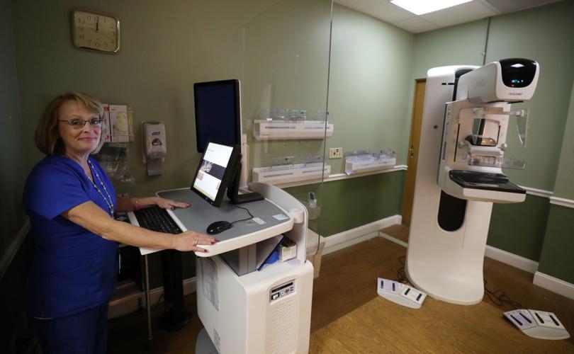
Diane Shoen, technologist for St. Mary’s, has administered mammograms for many years. She said a mammogram can be painful, and the new technology is no different. However, the quality of the image is better.
“They are in compression a little bit longer with the 3-D,” she said. “But just seconds, under 5 seconds.”
Along with the extra time in the machine, patients are required to hold their breath during the procedure because the equipment is extremely sensitive to motion, Shoen said.
According to Hologic’s guidelines, the maker of the 3-D mammography system at St. Mary's, the procedure can increase breast cancer detection by 20 percent, as well as a 15 percent decrease in false-positive rates.
Early detection of breast cancer is invaluable. The American Cancer Society recommends women consider a mammogram from ages 40 to 44, but begin yearly mammograms by age 45. She can consider exams every two years starting at age 55.
“If we can get the cancer at stage zero or one, we can have up to 95 percent cure rate,” Roubein said.
Contact Donnette Beckett at (217) 421-6983. Follow her on Twitter: @donnettebHR

Donnette Beckett
"Together Decatur" Columnist and Food/Drink Reporter


